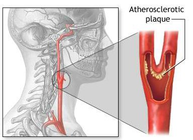
3D Plane-wave ultrasound matrix transducer for carotid artery diagnosis
Publications
- A Tiled Ultrasound Matrix Transducer for Volumetric Imaging of the Carotid Artery
dos Santos, Djalma Simões; Fool, Fabian; Mozaffarzadeh, Moein; Shabanimotlagh, Maysam; Noothout, Emile; Kim, Taehoon; Rozsa, Nuriel; Vos, Hendrik J.; Bosch, Johan G.; Pertijs, Michiel A. P.; Verweij, Martin D.; de Jong, Nico;
Sensors,
Volume 22, Issue 24, pp. 1--23, 2022. DOI: 10.3390/s22249799
Abstract: ...
High frame rate three-dimensional (3D) ultrasound imaging would offer excellent possibilities for the accurate assessment of carotid artery diseases. This calls for a matrix transducer with a large aperture and a vast number of elements. Such a matrix transducer should be interfaced with an application-specific integrated circuit (ASIC) for channel reduction. However, the fabrication of such a transducer integrated with one very large ASIC is very challenging and expensive. In this study, we develop a prototype matrix transducer mounted on top of multiple identical ASICs in a tiled configuration. The matrix was designed to have 7680 piezoelectric elements with a pitch of 300 μm × 150 μm integrated with an array of 8 × 1 tiled ASICs. The performance of the prototype is characterized by a series of measurements. The transducer exhibits a uniform behavior with the majority of the elements working within the −6 dB sensitivity range. In transmit, the individual elements show a center frequency of 7.5 MHz, a −6 dB bandwidth of 45%, and a transmit efficiency of 30 Pa/V at 200 mm. In receive, the dynamic range is 81 dB, and the minimum detectable pressure is 60 Pa per element. To demonstrate the imaging capabilities, we acquired 3D images using a commercial wire phantom.
document - Automated Characterization of Matrix Transducer Arrays using the Verasonics Imaging System
Djalma Simoes dos Santos; Fabian Fool; Taehoon Kim; Emile Noothout; Nuriel Rozsa; Hendrik J. Vos; Johan G. Bosch; Michiel A. P. Pertijs; Martin D. Verweij; Nico de Jong;
In Proc. IEEE International Ultrasonics Symposium (IUS),
2022. - Automated Characterization of Matrix Transducer Arrays using the Verasonics Imaging System
Djalma Simoes dos Santos; Fabian Fool; Taehoon Kim; Emile Noothout; Nuriel Rozsa; Hendrik J. Vos; Johan G. Bosch; Michiel A. P. Pertijs; Martin D. Verweij; Nico de Jong;
In Proc. IEEE International Ultrasonics Symposium (IUS),
2022. - Design of an Ultrasound Transceiver ASIC with a Switching-Artifact Reduction Technique for 3-D Carotid Artery Imaging
T. Kim; F. Fool; D. Simoes dos Santos; Z. Y. Chang; E. Noothout; H. J. Vos; J. G. Bosch; M. D. Verweij; N. de Jong; M. A. P. Pertijs;
Sensors,
Volume 21, Issue 1, pp. 150, January 2021. DOI: 10.3390/s21010150
Abstract: ...
This paper presents an ultrasound transceiver application-specific integrated circuit (ASIC) directly integrated with an array of 12 × 80 piezoelectric transducer elements to enable next-generation ultrasound probes for 3D carotid artery imaging. The ASIC, implemented in a 0.18 µm high-voltage Bipolar-CMOS-DMOS (HV BCD) process, adopted a programmable switch matrix that allowed selected transducer elements in each row to be connected to a transmit and receive channel of an imaging system. This made the probe operate like an electronically translatable linear array, allowing large-aperture matrix arrays to be interfaced with a manageable number of system channels. This paper presents a second-generation ASIC that employed an improved switch design to minimize clock feedthrough and charge-injection effects of high-voltage metal–oxide–semiconductor field-effect transistors (HV MOSFETs), which in the first-generation ASIC caused parasitic transmissions and associated imaging artifacts. The proposed switch controller, implemented with cascaded non-overlapping clock generators, generated control signals with improved timing to mitigate the effects of these non-idealities. Both simulation results and electrical measurements showed a 20 dB reduction of the switching artifacts. In addition, an acoustic pulse-echo measurement successfully demonstrated a 20 dB reduction of imaging artifacts.
document - Integrated Transceivers for Emerging Medical Ultrasound Imaging Devices: A Review
C. Chen; M. Pertijs;
IEEE Open Journal of the Solid-State Circuits Society,
Volume 1, pp. 104-114, September 2021. DOI: 10.1109/OJSSCS.2021.3115398 - Receive/Transmit Aperture Selection for 3D Ultrasound Imaging with a 2D Matrix Transducer
M. Mozaffarzadeh; M. Soozande; F. Fool; M. A. P. Pertijs; H. J. Vos; M. D. Verweij; J. G. Bosch; N. de Jong;
MDPI Applied Sciences,
Volume 10, Issue 15, July 2020. DOI: 10.3390/app10155300
Abstract: ...
Recently, we realized a prototype matrix transducer consisting of 48 rows of 80 elements on top of a tiled set of Application Specific Integrated Circuits (ASICs) implementing a row-level control connecting one transmit/receive channel to an arbitrary subset of elements per row. A fully sampled array data acquisition is implemented by a column-by-column (CBC) imaging scheme (80 transmit-receive shots) which achieves 250 volumes/second (V/s) at a pulse repetition frequency of 20 kHz. However, for several clinical applications such as carotid pulse wave imaging (CPWI), a volume rate of 1000 per second is needed. This allows only 20 transmit-receive shots per 3D image. In this study, we propose a shifting aperture scheme and investigate the effects of receive/transmit aperture size and aperture shifting step in the elevation direction. The row-level circuit is used to interconnect elements of a receive aperture in the elevation (row) direction. An angular weighting method is used to suppress the grating lobes caused by the enlargement of the effective elevation pitch of the array, as a result of element interconnection in the elevation direction. The effective aperture size, level of grating lobes, and resolution/sidelobes are used to select suitable reception/transmission parameters. Based on our assessment, the proposed imaging sequence is a full transmission (all 80 elements excited at the same time), a receive aperture size of 5 and an aperture shifting step of 3. Numerical results obtained at depths of 10, 15, and 20 mm show that, compared to the fully sampled array, the 1000 V/s is achieved at the expense of, on average, about two times wider point spread function and 4 dB higher clutter level. The resulting grating lobes were at −27 dB. The proposed imaging sequence can be used for carotid pulse wave imaging to generate an informative 3D arterial stiffness map, for cardiovascular disease assessment. - A 12×80 Element Ultrasound Transceiver ASIC With Enhanced Charge Injection Performance for 3-D Carotid Artery Imaging
T. Kim; F. Fool; E. Kang; Z. Y. Chang; E. Noothout; J. G. Bosch; M. D. Verweij; N. de Jong; M. Pertijs;
In Proc. IEEE International Ultrasonics Symposium (IUS),
September 2020. abstract. - 3D high frame rate flow measurement using a prototype matrix transducer for carotid imaging
F. Fool; H. J. Vos; M. Shabanimotlagh; T. Kim; E. Kang; M. Pertijs; N. de Jong; M. D. Verweij;
In Proc. IEEE International Ultrasonics Symposium (IUS),
IEEE, pp. 1-4, October 2019. - A Reconfigurable Ultrasound Transceiver ASIC With 24 × 40 Elements for 3D Carotid Artery Imaging
E. Kang; Q. Ding; M. Shabanimotlagh; P. Kruizinga; Z. Y. Chang; E. Noothout; H. J. Vos; J. G. Bosch; M. D. Verweij; N. de Jong; M. A. P. Pertijs;
IEEE Journal of Solid-State Circuits,
Volume 53, Issue 7, pp. 2065-2075, July 2018. DOI: 10.1109/JSSC.2018.2820156
Abstract: ...
This paper presents an ultrasound transceiver application-specific integrated circuit (ASIC) designed for 3-D ultrasonic imaging of the carotid artery. This application calls for an array of thousands of ultrasonic transducer elements, far exceeding the number of channels of conventional imaging systems. The 3.6 x 6.8 mm² ASIC interfaces a piezo-electric transducer (PZT) array of 24 x 40 elements, directly integrated on top of the ASIC, to an imaging system using only 24 transmit and receive channels. Multiple ASICs can be tiled together to form an even bigger array. The ASIC, implemented in a 0.18 μm high-voltage (HV) BCD process, consists of a reconfigurable switch matrix and row-level receive circuits. Each element is associated with a compact bootstrapped HV transmit switch, an isolation switch for the receive circuits and programmable logic that enables a variety of imaging modes. Electrical and acoustic experiments successfully demonstrate the functionality of the ASIC. In addition, the ASIC has been successfully used in a 3-D imaging experiment. - A Reconfigurable 24 × 40 Element Transceiver ASIC for Compact 3D Medical Ultrasound Probes
E. Kang; Q. Ding; M. Shabanimotlagh; P. Kruizinga; Z. Y. Chang; E. Noothout; H. J. Vos; J. G. Bosch; M. D. Verweij; N. de Jong; M. A. P. Pertijs;
In Proc. European Solid-State Circuits Conference (ESSCIRC),
IEEE, pp. 211-214, September 2017. - Towards 3D ultrasound imaging of the carotid artery using a programmable and tileable matrix array
P. Kruizinga; E. Kang; M. Shabanimotlagh; Q. Ding; E. Noothout; Z. Y. Chang; H. J. Vos; J. G. Bosch; M. D. Verweij; M. A. P. Pertijs; N. de Jong;
In Proc. IEEE International Ultrasonics Symposium (IUS),
IEEE, pp. 1-3, September 2017. DOI: 10.1109/ULTSYM.2017.8091570
Abstract: ...
Accurate assessment of carotid artery disease by measuring blood flow, plaque deformation and pulse wave velocity using ultrasound imaging requires 3D information. Additionally, the volume rates should be high enough (> 1 kHz) to capture the full range of these fast transient phenomena. For this purpose, we have built a programmable, tileable matrix array that is capable of providing 3D ultrasound imaging at such volume rates. This array contains an application-specific integrated circuit (ASIC) right beneath the acoustic piezo-stack. The ASIC enables fast programmable switching between various configurations of elements connected to the acquisition system via a number of channels far smaller than the number of transducer elements. This design also allows for expanding the footprint by tiling several of these arrays together into one large array. We explain the working principles and show the first basic imaging results of a 2-by-1 tiled array. - Optimizing the directivity of piezoelectric matrix transducer elements mounted on an ASIC
M. Shabanimotlagh; S. Raghunathan; V. Daeichin; P. Kruizinga; H. J. Vos; M. A. P. Pertijs; J. G. Bosch; N. de Jong; M. D. Verweij;
In Proc. IEEE International Ultrasonics Symposium (IUS),
IEEE, pp. 1-4, September 2017. DOI: 10.1109/ULTSYM.2017.8091752
Abstract: ...
Over the last decade, clinical studies show a strong interest in real-time 3D imaging. This calls for ultrasound probes with high-element-count 2D matrix transducer arrays. These may be interfaced to an imaging system using an in-probe Application Specific Integrated Circuit (ASIC) that takes care of signal amplification, element switching, sub-array beamforming, etc. Since the ASIC is made from silicon and is mounted directly behind the transducer elements, it can acoustically be regarded as a rigid plate that can sustain traveling lateral waves. These waves lead to acoustical cross-talk between the elements, and results in extra peaks in the directivity pattern. We propose two solutions to this problem, based on numerical simulations. One approach is to decrease the phase velocity in the silicon by reducing the silicon thickness and absorbing the energy using a proper backing material. Another solution is to disturb the waves inside the silicon plate by sub-dicing the back-side of the ASIC. We conclude that both solutions can be used to improve the directivity pattern. - The role of sub-dicing in the acoustical design of an ultrasound matrix transducer for carotid arteries imaging
M. Shabanimotlagh; J. Janjic; S. Raghunathan; M. A. P. Pertijs; N. de Jong; M. Verweij;
In Proc. IEEE International Ultrasonics Symposium (IUS),
IEEE, pp. 1‒4, September 2016. DOI: 10.1109/ultsym.2016.7728470
Abstract: ...
Accurate diagnostics of stenosis and blood flow distribution in carotid arteries requires transducers capable of producing 3D volume images with high frame rate for real time imaging. In the process of designing a matrix probe, an important goal is to realize the acoustic stack with high sensitivity and bandwidth. In this study, we employ a finite element analysis to evaluate the effect of sub-dicing on the performance of an acoustic stack in a piezoelectric matrix array. The array is integrated with an Application Specific Integrated Circuit (ASIC), which performs the task of signal amplification and efficient data reduction. The results show that two sub-dicing cuts can improve the sensitivity by 40%, bandwidth by 20%, and reduce the ringing time by 43%, which are all desired for improving the image quality.
BibTeX support